Introduction: What Is Royal Radiology?
At Royal Health, royal radiology means precise, clinically justified, and patient-centered medical imaging.
It is not about excessive scanning—it is about choosing the right modality, using the lowest effective exposure, and interpreting images within a clinical context.
Radiology plays a central role in modern healthcare, supporting diagnosis, treatment planning, and disease monitoring across nearly all medical specialties.
The Role of Radiology in Modern Medicine
Medical imaging allows clinicians to:
-
Visualize internal organs and structures non-invasively
-
Detect disease early, often before symptoms appear
-
Guide interventions and monitor treatment response
-
Improve diagnostic accuracy and patient outcomes
According to the World Health Organization (WHO), diagnostic imaging is essential for effective health systems worldwide.
https://www.who.int/health-topics/diagnostic-imaging
Core Imaging Modalities in Royal Radiology
Royal radiology relies on selecting the most appropriate imaging technique based on the clinical question.
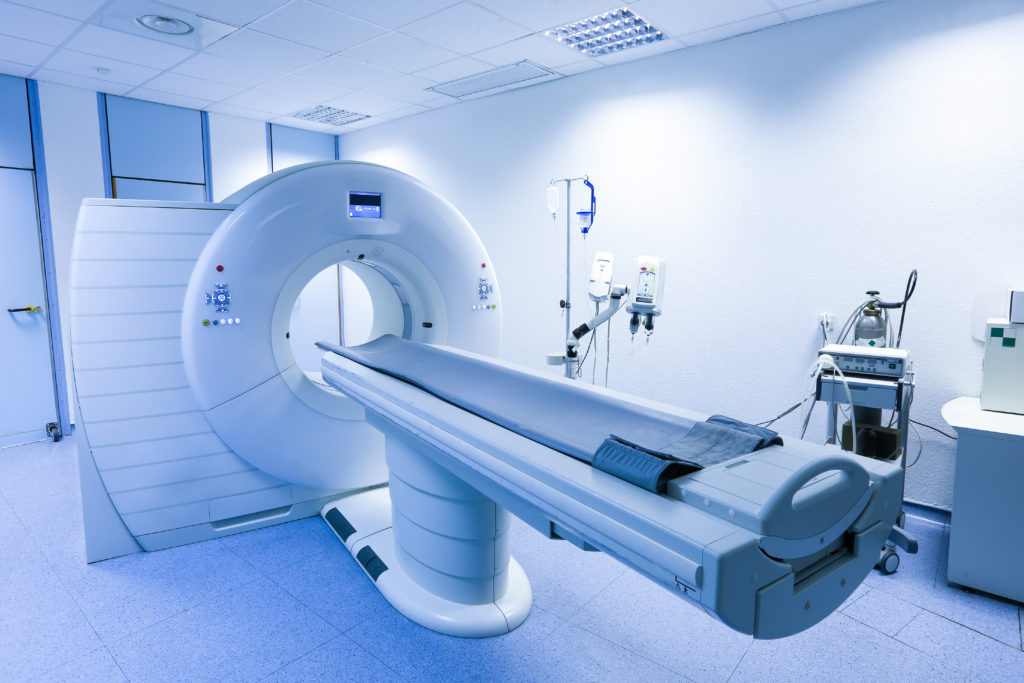
Computed Tomography (CT Scan)
What Is CT?
CT uses X-rays combined with computer processing to create detailed cross-sectional images of the body.
Clinical Uses
-
Trauma and emergency assessment
-
Brain hemorrhage and stroke evaluation
-
Chest and abdominal imaging
-
Cancer detection and staging
Safety and Evidence
CT scans involve ionizing radiation, but modern scanners use dose-optimization techniques to minimize exposure.
The Radiological Society of North America (RSNA) confirms CT as a highly valuable, evidence-based diagnostic tool when used appropriately.
https://www.radiologyinfo.org/en/info/bodyct
Magnetic Resonance Imaging (MRI)
What Is MRI?
MRI uses strong magnetic fields and radio waves, not radiation, to produce high-resolution images.
Clinical Uses
-
Brain and spinal cord disorders
-
Joint and soft tissue injuries
-
Cardiac and vascular imaging
-
Tumor characterization
Safety and Evidence
MRI is considered very safe but requires screening for metal implants or devices.
The National Institutes of Health (NIH) recognizes MRI as a cornerstone of advanced diagnostic imaging.
https://www.ncbi.nlm.nih.gov/books/NBK441998/
X-Ray Imaging
What Is X-Ray?
X-ray imaging uses low doses of ionizing radiation to visualize dense structures, especially bones and lungs.
Clinical Uses
-
Bone fractures and joint disorders
-
Chest infections and lung disease
-
Dental and orthopedic assessment
Safety and Evidence
X-rays involve minimal radiation exposure and remain one of the most widely used diagnostic tools globally.
The American College of Radiology (ACR) supports X-ray imaging as safe and effective when medically indicated.
https://www.acr.org/Clinical-Resources/Radiology-Safety
Ultrasound Imaging
What Is Ultrasound?
Ultrasound uses high-frequency sound waves, not radiation, to create real-time images.
Clinical Uses
-
Pregnancy and fetal assessment
-
Abdominal and pelvic organs
-
Thyroid, breast, and vascular imaging
-
Guidance for procedures
Safety and Evidence
Ultrasound is considered extremely safe and is often the first-line imaging tool, especially in children and pregnant patients.
The FDA and WHO both recognize diagnostic ultrasound as safe when used appropriately.
https://www.fda.gov/radiation-emitting-products/medical-imaging/ultrasound-imaging
What Makes Radiology “Royal”?
Royal radiology is defined by four principles:
1. Clinical Justification
Every scan answers a specific medical question.
2. Modality Optimization
The right test is chosen—CT, MRI, X-ray, or ultrasound—based on diagnostic need and safety.
3. Radiation Protection
When radiation is used, it follows the ALARA principle (As Low As Reasonably Achievable).
International guidelines from the International Atomic Energy Agency (IAEA) emphasize radiation safety.
https://www.iaea.org/topics/radiation-protection-of-patients
4. Expert Interpretation
Images are interpreted by trained radiologists and correlated with:
-
Clinical findings
-
Laboratory results
-
Patient history
Radiology without proper interpretation loses its value.
When Imaging Is NOT Royal (And Not Safe)
Imaging becomes inappropriate when:
-
Performed without clinical indication
-
Repeated unnecessarily
-
Used as a replacement for medical evaluation
-
Chosen without considering radiation exposure
The Choosing Wisely Campaign warns against unnecessary imaging.
https://www.choosingwisely.org
Royal Radiology and Preventive Health
Advanced imaging contributes to:
-
Early cancer detection
-
Cardiovascular risk assessment
-
Musculoskeletal degeneration monitoring
-
Organ health evaluation
Harvard Medical School highlights imaging as a key component of preventive and precision medicine.
https://www.health.harvard.edu
Patient Preparation for Royal Imaging
-
Follow fasting or hydration instructions
-
Inform staff about pregnancy or implants
-
Bring prior imaging for comparison
-
Use accredited radiology centers
Proper preparation improves image quality and diagnostic accuracy.
Final Thoughts: Royal Radiology Is Precision, Not Excess
Royal radiology is about intelligent imaging, not more imaging.
When CT, MRI, X-ray, and ultrasound are:
-
Clinically justified
-
Scientifically supported
-
Safely performed
-
Expertly interpreted
They become powerful tools for early detection, accurate diagnosis, and long-term health protection.
Dr. Ibrahem Abdelghany
Royal Health – Evidence-Based Wellness